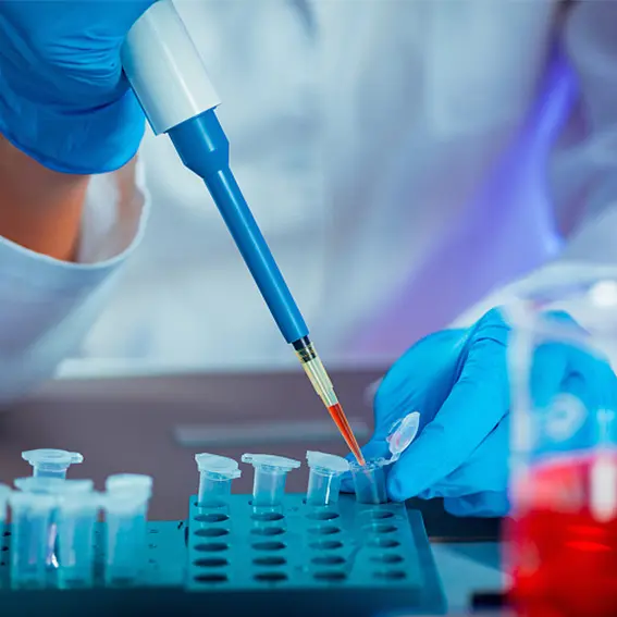
Real-time RT-PCR laboratories are now central to global public-health monitoring of respiratory pathogens. By amplifying trace nucleic-acid signatures in hours—or, with full automation, in minutes—these labs provide the sensitivity, specificity, and speed needed for large-scale screening and variant tracking. Yet performance is never just about the thermocycler; it depends on a carefully orchestrated workflow that merges biosafety, quality assurance, assay design, and robust data analytics. The guide below walks through the key components of a modern PCR facility, drawing on open-access resources from public universities and government agencies.
1. Facility Layout and Biosafety
A two-room layout—“clean” pre-amplification area and “post-amp” analysis area—remains the gold standard for contamination control. Core biosafety measures include directional airflow, hands-free doors, and dedicated PPE to prevent amplicon carry-over. Current U.S. CDC guidance recommends Biosafety Level 2 (BSL-2) infrastructure, plus an activity-specific risk assessment that documents engineering controls and personal protective equipment for each workflow step.
2. Sample Receipt and Pre-Analytical Workflow
Incoming respiratory swabs, saliva pools, or environmental surface wipes should be logged with a barcode LIMS that timestamps chain-of-custody events. Government guidance stresses that specimens be processed within four days of collection (ideally within 24 h) to maximize RNA integrity. Inactivation buffers or heat-inactivation steps are added here—not later—to protect downstream staff and instruments.
3. Automated Nucleic-Acid Extraction
High-throughput labs typically load 96- or 384-well extraction plates on liquid-handling robots to isolate RNA in under 30 min. University core facilities report that magnetic-bead platforms coupled with HEPA-filtered biosafety cabinets yield the most consistent recovery across respiratory sample types—while also freeing analysts to prepare the next batch.
4. Assay Design and Validation
Primer-probe sets should amplify 70–150 bp targets, with 40–60 % GC content, no homopolymers >4 nt, and melting temperatures within 1–2 °C of one another. Exon–exon junction spanning is preferred for RNA assays to eliminate genomic DNA noise. A validated limit of quantification ≤50 copies/µg gDNA—confirmed across three reagent lots—meets current federal guidance for molecular biodistribution tests.
5. Quality Assurance / Quality Control (QA/QC)
Written QA/QC plans should define positive, negative, and inhibition controls for every plate, plus acceptance criteria for Ct drift (<0.5 cycle run-to-run). U.S. EPA manuals and university core laboratories both emphasize quarterly proficiency panels and yearly thermocycler recalibration. Publishing the full QC dataset is a powerful trust signal to clients and regulators alike.
6. Data Analysis and Ct Interpretation
Automated baseline subtraction and thresholding reduce analyst bias. Labs commonly flag Ct > 38 as “trace detection” requiring repeat extraction, while values between 18-32 indicate ample template. Cornell University’s diagnostic center notes that reporting should focus on “detected” vs. “not detected,” avoiding over-interpretation of marginal cycles.
7. Troubleshooting Common Pitfalls
Frequent failure modes—primer-dimer artifacts, low fidelity polymerase errors, excess Mg²⁺—are best resolved by decreasing cycle count, shortening extension times, or swapping in a higher-fidelity enzyme. Comprehensive troubleshooting matrices from academic and government sources provide step-by-step fixes for each anomaly.
8. Contamination Control Strategies
Physical separation alone is insufficient. Labs add UV-C crosslinking cycles, bleach footbaths, and single-use consumables to further cut false positives. A “unidirectional flow” policy—people, plates, and pipettes move forward only—has proven to eliminate >95 % of amplicon contamination events in public-health pilot programs.
9. Scaling Up : Robotics and Parallel Workflows
For regional surveillance, capacity often jumps from 500 to >5 000 reactions per shift by integrating plate stackers, automated sealers, and multiplex chemistries that distinguish multiple respiratory targets in one well. University genomics cores illustrate how dual-plate thermocyclers and on-the-fly barcode parsing keep turnaround times under six hours—even when sample volume spikes.
10. Workforce Development and Continuing Education
Sustainable performance depends on a trained workforce. The CDC’s e-learning modules in molecular biology, along with on-site instrument certification from core facilities, ensure that analysts keep pace with protocol updates, software patches, and biosafety revisions. Many labs schedule quarterly refreshers and require a perfect score on blinded mock panels before recertification.
A high-efficiency PCR laboratory is far more than machines and reagents; it is an ecosystem of rigorously documented workflows, transparent quality metrics, and people empowered by continuous training. By adopting evidence-based biosafety practices, sharing full QC data, and automating wherever practical, laboratories can deliver rapid, reliable results that underpin large-scale respiratory-pathogen surveillance—without compromising safety, accuracy, or public confidence.